Zapytaj o cenę
Twoje dane kontaktowe
Prosimy o wypełnienie formularza oraz ewentualne wybranie (kliknięcie) interesujących
Państwa ultrasonografów i głowic z poniższego wykazu.
Wybierz aparaty
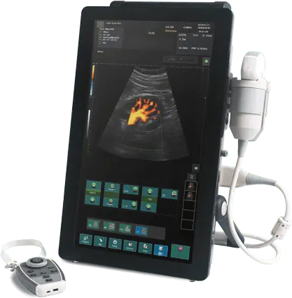
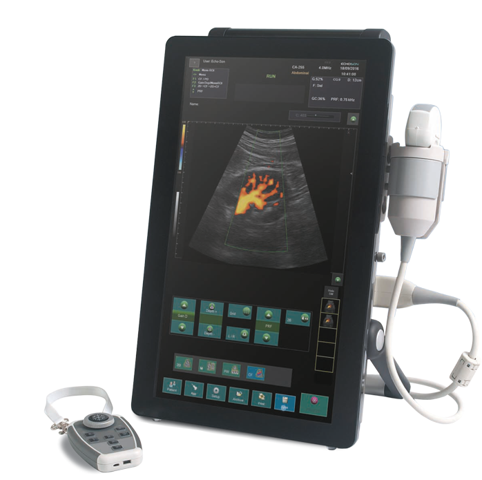
ALBIT Naczyniowy/ Anestezja
ALBIT Ortopedia/ MSK / Rehabilitacja
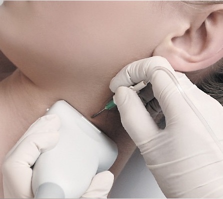
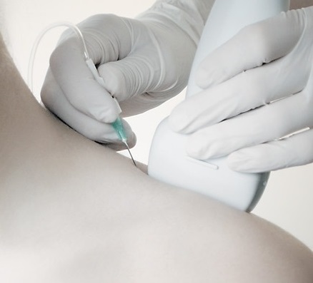
ALBIT Endokrynologia / Biopsja
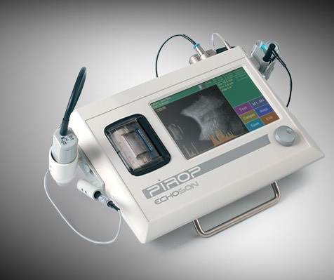
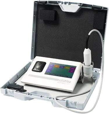
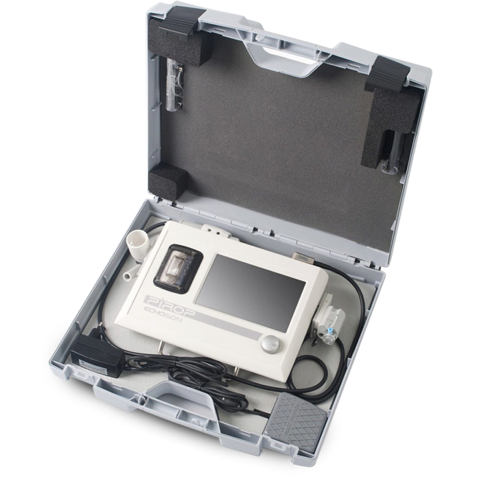
PIROP Biometr okulistyczny
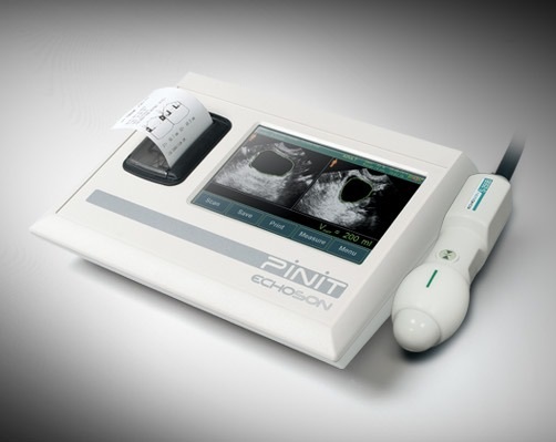
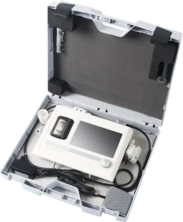
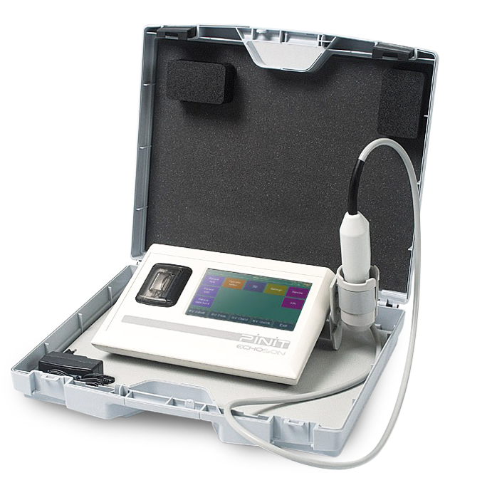
PINIT Skaner pęcherza
Wybierz głowice
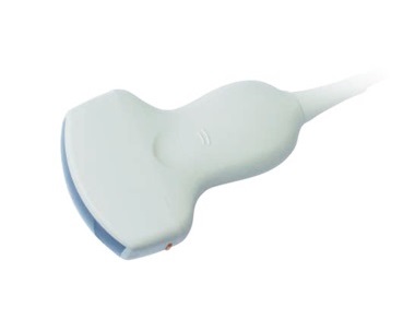
Convex: CA-255/R60
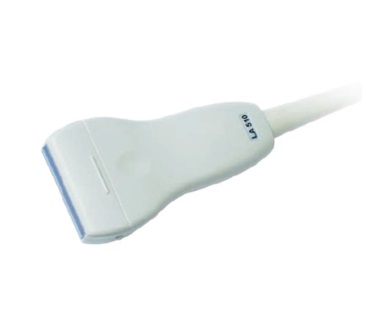
Liniowa: LA-510/L40
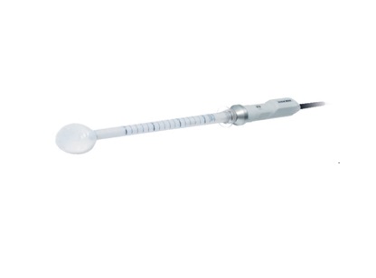
Anorektalna 360st : R-510/R8
Bląd! Wiadomość nie została wysłana! Proszę o wypełnienie wszystkich niezbędnych pól
Dziękuję! Wiadomość została wysłana
Wypełnienie powyższego formularza jest równoznaczne z wyrażeniem zgody na umieszczenie danych
osobowych w bazie firmy Echo-Son SA i wykorzystywaniem ich zgodnie z ustawą o ochronie danych osobowych. Dane te
będą wykorzystywane WYŁĄCZNIE przez firmę Echo-Son SA do celów kontaktów marketingowych/handlowych.
* pole wymagane

Arab Health 2024
Międzynarodowe
Targi Medyczne
Firma Echo-Son S.A. w dniach 29 stycznia – 2 lutego 2024 r. wzięła udział w największych na Bliskim Wschodzie Targach Medycznych Arab Health 2024 (Dubaj – ZEA). Zapraszamy do obejrzenie relacji z tego wydarzenia

tryb 3D
(opcja)
(opcja)

dotykowy
ekran
ekran

zdalny sterownik
ręczny
ręczny

stojak-wózek


Filozofią działania
Echo-Son S.A.
Echo-Son S.A.
jest dążenie do zapewnienia pełnej satysfakcji Klientów przez tworzenie aparatury ultradźwiękowej najwyższej jakości, spełniającej ich wymagania i wyprzedzającej oczekiwania, oraz zapewniając im jasno sformułowane zasady współpracy, profesjonalną i miłą obsługę.

Echo-Son
Wiodący polski producent ultrasonografów
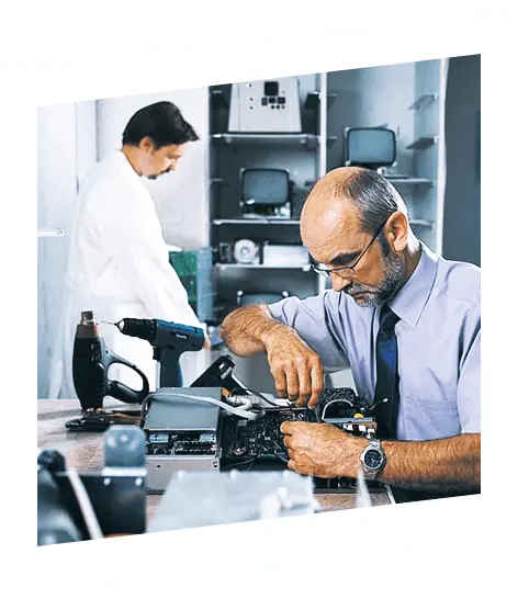
W obecnej formie organizacyjno-prawnej Zakład Doświadczalny Echo-Son S.A. powstał 30 lat temu w 1993 roku. Jesteśmy małą firmą, aktualnie w Echo-Son SA zatrudnionych jest około 40 osób.
Od 1993 roku Echo-Son SA konstruuje i produkuje ultradźwiękową aparaturę do diagnostyki medycznej. Oferta firmy obejmuje szeroką gamę ultrasonografów spełniających najwyższe standardy i wymagania. Ultrasonografy Echo-Son charakteryzują się dużą uniwersalnością i nowoczesnością przez zastosowanie w nich m.in. cyfrowej technologii emisji i odbioru fali ultradźwiękowej oraz cyfrowego zobrazowania przepływu krwi – Color-Doppler. Głównym akcjonariuszem Echo-Son S.A. jest Instytut Podstawowych Problemów Techniki Polskiej Akademi Nauk.
Echo-Son posiada certyfikat ISO-9001 (ISO-13485) od 2000 roku, oraz certyfikat CE (MDD-93/42/EEC) od 2002 roku, jeszcze przed przystąpieniem Polski do Unii Europejskiej.
Echo-Son eksportuje produkowane ultrasonografy do wielu krajów na całym świecie w tym do krajów Unii Europejskiej, Bliskiego i Dalekiego Wschodu, Północnej Afryki i Ameryki Łacińskiej.
Zapraszamy
Zespół Echo-Son
ZOBACZ WIĘCEJ
30
lat
na rynku
na rynku
59
krajów
eksportu
eksportu
30
inzynierów
specialistów
specialistów